

PB 24082
Description
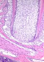
Late stage development of the first molar root. Root formation is still actively ongoing with deposition of dentine matrix within the pre-dentine zone (pre-dentine appears pale in comparison to the mineralised dentine) In the most apical region close to Hertwig`s root sheath, differentiation of odontoblasts is evident due to the transition from low cuboidal to cylidrical cell morphology with apically placed nuclei. Odontoblast differentiation occurs from ectomesenchymal neural crest derived precursor cells.
The pulpal cavity is richly vascularised and composed of immature fibroblast-like cells situated in a very loose connective tissue matrix.
The root surface borders on an immature periodontal ligament composed of irregularly organised fibroblasts within a richly vascularised collagenous meshwork. In the upper left part of the image immature alveolar bone is present in which the bony trabeculae are lined by active osteoblasts.
In the lower left part of the picture the mandibular canal and the mandibular nerve can be seen.
MPATH / Pathology
MPATH 458 - normal
Gene
sex
Male
strain
C57BL/6 CRL
organism
Mouse
EMAP / Embryonic stage, tissue or post-natal age:
genotype status
Wild-type
genetic manipulation
None
MA / Anatomical Site
MA 1602 - lower jaw molar
Designated Allele Name
Experimental Manipulation
Further info
NOTE: Not all terms are currently shared between Pathbase and the above databases, some searches may not produce returns, in which case users should use synonyms or more inclusive text terms to search manually!
Copyright
This image remains the property of the originating Institution and should not be modified, reproduced or disseminated without the express permission of the submitter.